Chiari Malformation und Syringomyelie
Weiterhin finden Sie hier alle weiteren wichtigen Informationen Rund um diese Themengebiete.
Umgang
Psyche
Grundsätzliches über Syringomyelie, Hydromyelie und (Arnold)-Chiari Malformation
Q07.0 Chiari Malformation
G95.0 Syringomyelie
(keine vollständige Aufzählung):
- Arachnopathie
- Berührungsempfindlichkeit der Gliedmaßen und anderer Körperregionen
- Depressionen oder Verstimmungen
- Ermüdungszustände, allgemeine Kraftlosigkeit, Neigung zu schneller Erschöpfung
- Faszikulationen (unwillkürliche Zuckungen einzelner Muskelfaserbündel)
- Fehlregulationsbedingte Durchblutungsstörungen
- Gangunsicherheit
- gesteigerte Hitze- oder Kälteempfindlichkeit (bzw. Unempflichkeiten)
- Inkontinenz von Blase und Darm bis hin zur Lähmung des Blasen- bzw. Schließmuskels
- Lichtempfindlichkeit
- Missempfindungen in den Gliedmaßen wie Kribbeln oder stechen
- Muskelatrophie
- Neurogene Bauchschmerzen
- partieller Muskelschwund
- Schlafapnoe
- Schluckbeschwerden, Zungendysfunktion
- Schwindel und Koordinationsstörungen
- Sensibilitätsstörungen wie Hitzeunempfindlichkeit einzelner Hautpartien der Gliedmaßen
- sexuelle Funktionsstörungen
- Skoliose
- spastische Lähmungen
- Starke scharfe, brennende oder dumpfe Schmerzen im Bereich von Schulter, Kopf, Nacken und in den Armen, migräneartige Kopfschmerzen
- Störung der Tiefensensibilität (die anzeigt, in welcher Stellung sich der Körper oder die Gliedmaßen befinden)
- Störungen des Lagesinns
- Störungen im Magen und Darmstrakt
- temporäre Gedächtnisstörungen (bzw. Wortfindungsstörungen)
- verlangsamte Wundheilung
- Verschlechterung des Sehvermögens
Mobilität:
Hilfsmittel
Weiterführende Links zur Erkrankung
Literaturtipps:
If these causes can be ruled out, one often speaks of hydromyelia (Greek: hydros = water) (to differentiate it from syringomyelia). Patients with hydromyelia can sometimes also suffer from severe pain and abnormal sensations, but the signs of the disease and the expansion of the cavity in the spinal cord do not increase further over time - they are not "progressive". The treatment of this often congenital enlargement of the central spinal canal (central canal) is therefore also purely symptomatic. You don't have to interrupt a progressive physical disease process here to stop the disease.
Arnold Chiari malformation (malformation)
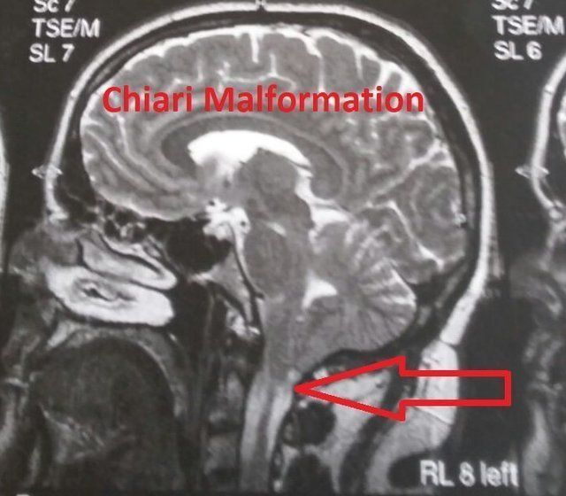
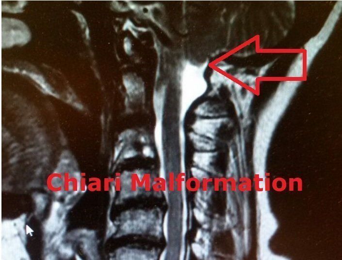
Arnold-Chiari malformation or just Chiari malformation is a malformation of the transition between the occiput and the cervical spine. Essentially, parts of the cerebellum (tonsils) shift into the occipital cavity, which affects the circulation of the cerebral water in this area.
Due to the different changes that can develop in this malformation during the embryonic period, different types are distinguished.
The Chiari malformation is one of the most common embryonic development disorders (8 per 100,000) and forms in the 6th to 10th grade. Week of pregnancy. However, it is not inherited. ACM is innate. Chronically increased intracranial pressure can also lead to a displacement of the cerebellar tonsils into the occipital cavity, which can lead to symptoms similar to those of ACM. The symptoms usually begin in childhood and adolescence, but it can also take 30-40 symptom-free years before those affected notice symptoms.
Type I Chiari malformation is almost always present.
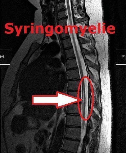
In type I, the cerebellar tonsils are shifted into the occipital opening, which is often narrow in the bones. In addition, the spider's skin (arachnoidea), as one of the shells of the brain, can be so fused that the circulation of cerebral and spinal fluid is hindered. This circulatory disorder can be the cause of syringomyelia (cavity formation in the spinal cord, see there), which can spread over the entire spinal cord and thus cause symptoms throughout the body.
In type II there is a slight to severe displacement of the cerebellum combined with a malformation of the brain stem. Nerve cells die in the displaced brain tissue. In about 80% of those affected, the brain chambers (ventricles), which form the cerebral and nerve water, are abnormally changed, so that the cerebral water is stagnated. Such a jam leads to an expansion of the brain chambers (hydrocephalus = head of water). If the cranial sutures are not yet closed, as in small children, the skull expands in volume and appears too large compared to the small body.
Type III is extremely rare, in which, in addition to the changes in type II, the cerebellum and / or parts of the brain stem shift to the neck area. This tissue displacement can emerge if there is a developmental gap in the skull bone, known as an encephalocele. Children with ACM III are hardly viable due to the severe damage
Type IV is even rarer. It describes a genetically determined underdevelopment (hypoplasia) of the cerebellum with a smaller posterior fossa that is essentially filled with cerebral fluid.
The symptoms that occur in ACM depend on the type of Chiari malformation and whether there are additional diseases. These are the following symptoms: headache and neck pain
- Dizziness and unsteady gait, especially when looking up
- Sensory disturbances (e.g. tingling in the hands, sensory deficits)
- Incoordination
- Motor paralysis of the arms and legs
- flaccid paralysis
- spontaneous rhythmic eye movements (nystagmus)
- Speech disorders, especially in the sense of slurred speech
- Decreased coughing and swallowing reflex, difficulty swallowing
The aim of treatment is to eliminate the malformation and its effects. Treatment options consist primarily of a decompression operation with widening of the posterior fossa and the occipital orifice, the shrinking of growths of the cerebellum, the loosening of adhesions in the posterior fossa and an enlargement of the hard meninges in the head-neck junction. After a successful operation, in many cases an existing syrinx (cyst in the spinal canal, see there) regresses.
If there is a build-up of cerebral fluid in type II, a drainage tube (shunt) can be implanted in the fourth ventricle to drain excess liquor (cerebral fluid) from the cerebral ventricle. This regulates the resulting overpressure in the cranial brain area. In addition to this drainage, treatment as described above for type I is usually required.
With types III and IV, viability and operability are very much dependent on the circumstances of the individual case, so that no general statements can be made about treatment.
The diagnosis of a Chiari malformation is made with the help of magnetic resonance imaging (MRI, also magnetic resonance imaging). As a result, the changes characteristic of the Chiari malformation are clearly mapped. Additional findings, such as the presence of a syrinx, can also be determined in terms of position and extent. By using a contrast medium that is injected during the examination, pathological changes such as tumors can also be made visible. Computed tomography can show any existing bony malformations better than an MRI.
In order to visualize the circulation of the nerve water in the skull or in the spinal canal, specialized clinics are able to record the flow with a special MRT examination (see figure below). The pulsation of the nerve water is displayed with each heartbeat of the person being examined. In addition, through an exact analysis of very fine-grained MRI examinations (FIESTA, CISS), even the smallest adhesions of the spinal cord and its envelopes with impaired circulation of the nerve water can be recognized. Electrophysiological examinations such as SSEP (somatosensory evoked potentials) or MEP (motor evoked potentials) can sometimes be important for the differential diagnosis of sensory disturbances.
The removal of nerve fluid from the spinal canal (lumbar puncture) can also provide important information about other pathological changes in the central nervous system. However, if the occipital opening is blocked at the same time, it can be life-threatening and should generally be postponed until operative decompression has taken place.
With the help of special ultrasound examinations, a Chiari malformation can already be recognized in the unborn child in the uterus.
The disease descriptions were given with the kind permission of Prof. Dr. med. Steffen Rosahl was taken over by the website of the Helios Klinikum Erfurt.
Furthermore, we have an interview with a specialist in neurology - chief physician Mr. Dr. T. Graf - from the Bad Kötzing rehabilitation clinic.
As well as a contribution on the subject of "Pregnancy and Diving" by Prof. Dr.med. Steffen Rosahl from the Helios clinics in Erfurt.
Berichte
Fragebögen
Weiterführende Links:
-
Deutsche Schmerzgesellschaft e.V.
Diagnose - Symptome - Schmerzen bei Syringomyelie - Behandlung
-
biowellmed - Ihr Portal für Gesundheit
biowellmed - Ihr Portal für Gesundheit
-
Beobachter GESUNDHEITListenelement 1
Beobachter: News & Infos vom sozialsten Netzwerk der Schweiz
Wissen, was wichtig ist: Was sind die neusten Abzocker-Tricks? Welchen Firmen und Politikern muss man genau auf die Finger schauen? Hier erfahren Sie es.
-
ONMEDAListenelement 2
Onmeda.de steht für hochwertige, unabhängige Inhalte und Hilfestellungen rund um das Thema Gesundheit und Krankheit. Hier finden Sie Antworten auf Fragen zu allen wichtigen Krankheitsbildern, Symptomen, Medikamenten und Wirkstoffen. Außerdem bietet Onmeda hilfreiche Informationen zu Ihrem Arztbesuch, indem es über Behandlungen und Untersuchungen aufklärt. Die Inhalte sind genau recherchiert, auf dem aktuellen Stand von Wissenschaft und Forschung und verständlich erklärt.
-
Helios KlinikenListenelement 4
NeuroMedizin
Erkrankungen des Nervensystems können vielfältige Ursachen haben. Neuromediziner sind ständig auf der Suche nach diesen Ursachen: im Labor, in Krankenhäusern und Praxiseinrichtungen, in Arbeitsgruppen auf regionaler, nationaler und internationaler Ebene.
Thanks for your interest
Das klingt banal, beschreibt aber den Kern des Problems vieler Patienten.
(Prof. Dr. med. Uwe Max Mauer)
Leitender Oberstarzt der Neurchirurgie des Bundeswehrkrankenhaus Ulm


